Home
/ Foot And Leg Bones Diagram : Leg Ankle And Foot Bones Diagram Quizlet - Ankle & lower leg anatomy.
Foot And Leg Bones Diagram : Leg Ankle And Foot Bones Diagram Quizlet - Ankle & lower leg anatomy.
Foot And Leg Bones Diagram : Leg Ankle And Foot Bones Diagram Quizlet - Ankle & lower leg anatomy.. Bone diagram forehead (frontal bone) nose bones (nasals) cheek bone (zygoma) upper jaw (maxilla) lower jaw (mandible) breast bone (sternum) upper arm bone (humerus) lower arm bone (ulna) thigh bone (femur) collar bone (clavicle) toe bones (phalanges) ankle bones (tarsals) kneecap (patella) shin bone (tibia) calf bone (fibula) foot bones It extends from your knee joint upwards to the ankle joint downwards. This bone creates the lower portion of the ankle joint.; On the left side of the image, above the heel, you can see the delicate leg bone called the fibula. Having two separate bones instead of one connecting the foot to the leg gives the foot and leg extra balance and maneuverability.
The ankle consists of three bones attached by muscles, tendons, and ligaments that connect the foot to the leg. The ankle joint is both a synovial joint and a hinge joint. These bones articulate (connect) to the talus or ankle bone at the tibiotalar joint (ankle joint) allowing the foot to move up and down. The knee joint is the largest joint in the body and is primarily a hinge joint, although some sliding and rotation occur. Leg, ankle and foot bones.
Bones And Joints Of The Foot And Ankle Overview Footeducation from footeducation.com The bones of your leg and foot helped give you the ability to score that field goal. Leg, ankle and foot bones. The talus is held in place by the foot bones surrounding it and various ligaments. The smaller lateral bone of the lower leg. Knee, leg, and foot (overview) how many times have a layman's language and anatomy ever matched? The talus is the bone at the top of the foot. As these nerves descend toward the thighs, they form two networks of crossed nerves known as the lumbar plexus and sacral plexus. Is the outer and narrower of the 2 lower leg bones, extending from the knee to.
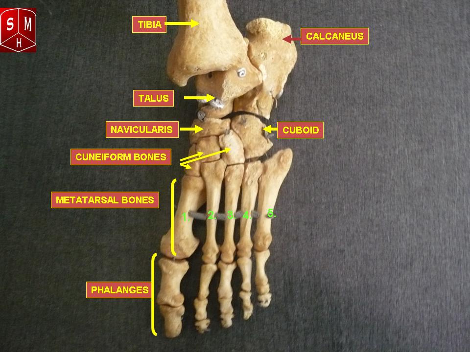
Tibia and fibula (long bones) the foot is connected to the body where the talus articulates with the tibia and fibula.
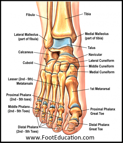
The ankle joint is both a synovial joint and a hinge joint. The medial, larger bone of the lower leg. Bones of the leg and foot, lower leg bone anatomy, leg bones anatomy, leg muscles, leg bones diagram, leg bone structure, leg anatomy muscles, parts of the lower leg. The ankle joint is the shock absorber of the foot. Having two separate bones instead of one connecting the foot to the leg gives the foot and leg extra balance and maneuverability. The calcaneus (heel bone) is the largest bone in the foot. Interestingly, no muscles attach to the talus. The talus is the bone at the top of the foot. The thigh bone or longest bone in the body. The talus bone supports the leg bones (tibia and fibula), forming the ankle. It connects with the tibia and fibula bones of the lower leg. The ankle joint or tibiotalar joint is formed where the top of the talus (the uppermost bone in the foot) and the tibia (shin bone) and fibula meet. In the lower leg are two bones called the tibia (shin bone) and the fibula.
Is the inner and larger of the 2 lower leg bones, extending from the knee to the ankle. Apart from 28 bones, 33 joints, muscles, ligaments, and about 100 foot tendons make the foot. Learn with flashcards, games, and more — for free. On the left side of the image, above the heel, you can see the delicate leg bone called the fibula. While we talk concerning leg anatomy worksheets, we've collected particular similar images to give you more ideas.
Foot Anatomy Bones Ligaments Muscles Tendons Arches And Skin from biologydictionary.net The bones of the leg and foot form part of the appendicular skeleton that supports the many muscles of the lower limbs. On the left side of the image, above the heel, you can see the delicate leg bone called the fibula. Leg, ankle and foot bones. These muscles work together to produce movements such as standing, walking, running, and jumping. Its made up of 4 bones; The talus bone supports the leg bones (tibia and fibula), forming the ankle. The seven tarsal bones are: These bones articulate (connect) to the talus or ankle bone at the tibiotalar joint (ankle joint) allowing the foot to move up and down.
The medial, larger bone of the lower leg.
Bones of the leg and foot, lower leg bone anatomy, leg bones anatomy, leg muscles, leg bones diagram, leg bone structure, leg anatomy muscles, parts of the lower leg. Helping the foot withstand the weight of the body whilst standing and in motion. The bones of the foot provide mechanical support for the soft tissues; Is the outer and narrower of the 2 lower leg bones, extending from the knee to. These muscles work together to produce movements such as standing, walking, running, and jumping. As these nerves descend toward the thighs, they form two networks of crossed nerves known as the lumbar plexus and sacral plexus. The talus is the bone at the top of the foot. Tibia and fibula (long bones) the foot is connected to the body where the talus articulates with the tibia and fibula. It forms the bottom of the ankle joint, articulating with the tibia and fibula (shin bones) and the top of the subtalar joint, articulating with the calcaneus (heel bone). The medial, larger bone of the lower leg. The talus is the highest foot bone. Its made up of 4 bones; The bones of the leg are the femur, tibia, fibula and patella.the foot bones shown in this diagram are the talus, navicular, cuneiform, cuboid, metatarsals and calcaneus.
It extends from your knee joint upwards to the ankle joint downwards. Let's review all of these bones one last time. When one looks at the anatomy of the foot, they would realize that the foot has a complex mechanical and structural architecture. Typically caused by trauma or injury to the foot or toe. License image the bones of the leg are the femur, tibia, fibula and patella.
Muscular Anatomy Muscle Anatomy Human Body Anatomy Body Anatomy from i.pinimg.com The lumbar plexus forms in the lower back from the merger of spinal nerves l1 through l4 while the. Knee, leg, and foot (overview) how many times have a layman's language and anatomy ever matched? The bones of the foot provide mechanical support for the soft tissues; Apart from 28 bones, 33 joints, muscles, ligaments, and about 100 foot tendons make the foot. #diagram and names of leg bones #diagram of foot and leg bones #diagram of leg bones #diagram of lower leg bones #diagram of the bones in your leg related posts of diagram of leg bones inside of arm muscle and bone The seven tarsal bones are: The talus is the bone at the top of the foot. They can be divided into three groups:
Learn with flashcards, games, and more — for free.
Learn with flashcards, games, and more — for free. Lower limb blood vessel coloring page, lower limb muscles anatomy worksheet and foot and ankle bones unlabeled are three of main things we want to present to you based on the gallery title. The talus is the bone at the top of the foot. The ankle is a joint that connects the lower leg to the foot. There are a whole range of structures e.g. Ankle & lower leg anatomy. Its main function is to allow for plantar flexion and dorsiflexion of the foot. The nerves of the leg and foot arise from spinal nerves connected to the spinal cord in the lower back and pelvis. The knee is a strong but flexible hinge joint. Sits over the front of the knee joint. Fpe medical review board a foot pain diagram is a great tool to help you work out what is causing your ankle and foot pain. They can be divided into three groups: The calcaneus (heel bone) is the largest bone in the foot.
